


4yr
Immunohistopathological Analysis of Immunoglobulin E-Positive Epidermal Dendritic Cells with House Dust Mite Antigens in Naturally Occurring Skin Lesions of Adult and Elderly Patients with Atopic Dermatitis
Source : https://www.mdpi.com/2296-3529/8/3/45/htm
The immunopathogenic role of house dust mite (HDM) allergens in the development of skin lesions in atopic dermatitis (AD) has not yet been precisely clarified. We immunohistopathologically evaluated the localization of immunoglobulin E (IgE)-positive epidermal dendritic cells with HDM antigens in the skin lesions of patients with IgE-allergic AD.
• Conclusion/Relevance: “We consider that the results of the present study demonstrated the crucial role of HDM [house dust mite] allergens in the immunopathogenesis of eczematous dermatitis in IgE-allergic AD [atopic dermatitis], in which the IgE-mediated delayed-type hypersensitivity reaction, together with IgE-bearing DCs [dendritic cells] (i.e., IDECs, LCs, and dermal inflammatory DCs), specific T cells, keratinocytes, and HDM antigens, may lead to spongiosis formation.
• In the current study, authors analyzed biopsy specimens from the skin lesions of six patients with IgE-allergic AD and HDM allergy, in addition to 11 control subjects having inflammatory skin disorders.
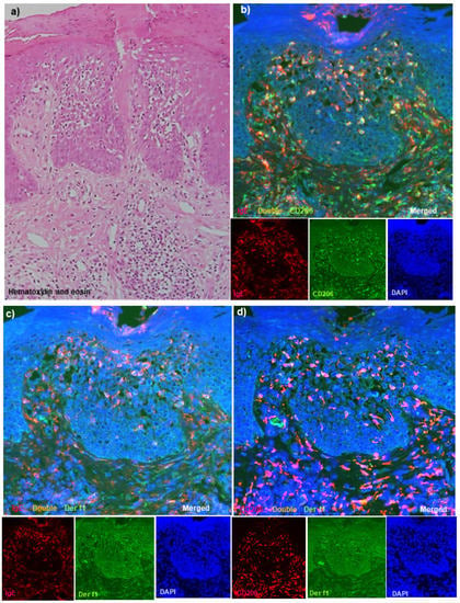
• Using double-immunofluorescence staining techniques in adult and elderly patients with IgE-allergic AD and HDM allergy, the investigators confirmed that IgE-bearing IDECs (CD11c+ and CD206+ cells) infiltrated and aggregated in the central area of the spongiotic epidermis in active lesions of chronic AD, along with T-cell infiltration.
• The authors wrote, “In the analysis of cell infiltration in the upper dermis, infiltrating double-positive IgE+ CD11c+ cells (i.e., IgE-bearing dermal inflammatory DCs) were mainly observed in the papillary and subpapillary dermis in the lesioned skin of patients with IgE-allergic AD; the numbers were significantly higher in patients with IgE-allergic AD than in the control subjects with non-eczematous inflammatory skin disorders and serum hyper-IgE.”
• Limitations of the current study included technical limitations of immunohistopathological analyses, no blind method for quantitative evaluations, and small AD sample sizes, as well as control cases. The investigators also did not include patients with non-IgE-allergic (intrinsic form) AD or enough patients with other types of eczematous disorders with spongiotic epidermis (e.g., allergic contact dermatitis) as study/control subjects.